

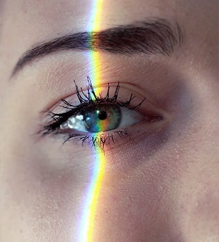
Learn More About Your Eyes & How They Work
When we perform a comprehensive eye exam, we take a look at several essential parts of your eye. Your eyes use complex systems to maintain its health and provide you with clear, comfortable vision.
At Family EyeCare Center Optometry, we believe that education is also a great way to help our patients manage and maintain their eye health, so we’re happy to answer any and all questions you have regarding your vision and your eye anatomy.
For clear, simple answers about your particular set of eyes, be sure to book an appointment with our team today!
The Front of Your Eyes
Conjunctiva
The front surface of your eyes and inner layer of your eyelids are covered by a thin membrane called the conjunctiva.
The conjunctiva secretes mucus that is used by your tear film across your eye, keeping them comfortable, hydrated, and protected from dust and dirt.
Cornea
The cornea is the front surface of your eye that covers your iris and pupil. The cornea has several different layers that work together to help focus light on your retina to deliver crisp, clear vision. These layers include:
- The epithelium
- Bowman’s layer
- Corneal stroma
- Descemet’s membrane
- The endothelium
Your cornea is responsible for 65% to 75% of your eye’s focusing power, while your eye’s lens takes on the rest of that responsibility.
Anterior Chamber
The anterior chamber is a space between your iris and cornea that includes a fluid called the aqueous humor. Your aqueous humor provides nutrients to your eyes and provides the pressure it needs to maintain its shape.
The aqueous humor flows through the anterior chamber to a spongy meshwork called the trabecular meshwork, draining out of your eye. However, blockages in this drainage system can cause high intraocular pressure, leading to a disease called open-angle glaucoma.
Iris
Your iris is the part of the eye that determines your eye’s color and controls your pupil’s size depending on how much light the pupil takes in. The iris is made up from connective tissues and muscles.
Lens
The lens is located right behind the iris and is responsible for fine-tuning the light that enters your eye to focus on your retina.

Inside Your Eyes
Vitreous Humor
The vitreous humor is a clear, gel-like substance that occupies the space between your lens and retina. If fluids or other substances enter or develop your vitreous humor, you may notice them as flashes or floaters.
Retina
The retina is a thin layer of tissue that lines the inside walls of your eyes. The retina houses millions of receptors that translate the light your eyes receive into neural signals for your brain to interpret.
Diseases like diabetic retinopathy can damage your retina over time, leading to vision loss.
Macula
The macula is the centermost part of your retina, responsible for providing the sharp vision you need to read, drive, and recognize faces. Your macula also houses a small depression in it’s center called the fovea, which is responsible for providing your sharpest vision.
Several diseases can damage your macula, including age-related macular degeneration and diabetic macular edema.
Optic Nerve
The optic nerve is a group of nerve fibres responsible for taking the information your retina collects to your brain. The connection point between the nerve and retina, called the optic disc, can be observed during a comprehensive eye exam to look for signs of diseases like glaucoma.
Book an Appointment Today
Family EyeCare Center Optometry is here to provide your family with all the information they need to make informed decisions about their eye health.
We’re ready to provide the care you need, and all you have to do is book an appointment today!
Come See What We’re All About
Visit Us Today
Find our practice on East Hamilton Drive right next to 7 Leaves Cafe. Parking is behind our building with accessible options available. Same day appointments available! Welcoming new patients.
- 338 E Hamilton Ave
- Campbell, CA 95008
Hours of Operation
- Monday: 9:00 AM – 6:00 PM
- Tuesday: 10:00 AM – 6:00 PM
- Wednesday: 8:30 AM – 6:00 PM
- Thursday: 9:30 AM – 6:00 PM
- Friday: 8:30 AM – 5:00 PM
- Saturday: 8:00 AM – 3:00 PM
- Sunday: Closed


Our Brands

Our Google Reviews

-
Total Vision Campbell
- 338 E Hamilton Ave
- Campbell, CA 95008
-
P: +14088662020
campbell@totalvisionllc.com